Lecture Notes, Biology 203, Human Sexuality and Reproduction
Introductory Material
- Anatomy and Physiology
- Definitions
- Anatomy: location, shape, size, appearance of structures
- Physiology: function of structures--how they work, when they work, in relation to each other
- Male reproductive tract anatomy and physiology
- Testis
- Sperm originate in wall of seminiferous tubules
- Cells near outer edge of wall differentiate into more mature cells closer to center of wall
- Nearly mature sperm are released into hollow center of tubule and travel to epididymis
- Interstitial cells between tubules produce testosterone, which promotes sperm maturation
- Epididymis
- Sperm continue to mature in epididymis
- Complete maturation of sperm requires about 45 days in the testis and about 18 days in the epididymis (about 63-65 days total)
- Mature sperm are composed of head (acrosome with enzymes, DNA), midpiece (mitochondria -> energy), and tail (motility)
- Vas deferens, ampulla of vas deferens
- Seminal vesicles
- Produce much of fluid in semen
- Nutritional chemicals: fructose, amino acids, lipids
- Prostate gland
- Secretes an alkaline fluid that enters urethra through pores in urethral walls
- Components: buffering chemicals, citrate, calcium, spermine
- Sperm are both viable and motile at pH 7-8.5, and begin to be less motile and viable below pH 7.0; since normal vaginal pH is 3.8-4.5, sperm must be protected from acidic pH by components of semen
- Bulbourethral gland (Cowper's gland) (secrete viscous alkaline fluid)
- Ejaculatory duct
- Urethra
- Semen components and normal characteristics
- Volume: 2.5 to 5 ml per ejaculation
- Sperm (from epididymis, testis)-- 50 to 150 million per ml (less than 20 million suggests infertility)
- Nutrients and fluid (from seminal vesicles)
- Protective, buffering chemicals (from prostate gland)
- Thick, alkaline fluid (from bulbourethral glands)
- Color: milky appearance (due to secretions from prostate)
- Motility: at least 60% active, showing good forward motion)
- pH: slightly alkaline (pH of 7.2 and 7.6 or 7.8)
- Specific gravity: 1.028 (slightly denser than water)
- Morphology: at least 80% normal
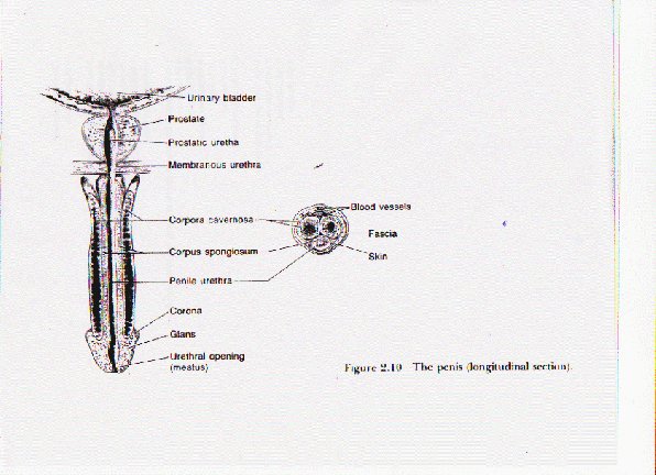
- Penis
- Shaft, glans, coronal ridge
- Erectile tissues: corpora cavernosa, corpus spongiosum
- Urethra passes through corpus spongiosum to urethral opening
-
- Nervous system control of erection and ejaculation
- Brain communicates sexual stimulus to erection reflex center in sacral part of spinal cord
- Spinal cord nerves send signals to erectile tissue in penis, arteries dilate, blood pools in erectile tissues, leading to erection
- Spinal cord nerves send signals that result in contraction of inner structures (epididymis, vas deferens, seminal vesicles, prostate)
- Semen washes past bulbourethral glands into the lower urethra (emission); sensation of urethral filling is returned to erection complex
- Spinal cord nerves send signals that result in contraction of muscles at base of penis, leading to rhythmic contractions (ejaculation)
- Female reproductive tract anatomy and physiology
- Production of ova
- A female embryo, at two month postconception (about seven months before birth): ovary has 600,000 germ cells in her ovaries(ancestors of eggs or ova)
- By 5 months postconception, the fetus has 6-7 million germ cells, which will develop into 5 million primary oocytes (peak number of oocytes for her lifetime)
- At the time of birth, 700,000 to 2 million primary oocytes will remain in the ovaries
- By puberty, 40,000 to 400,000 are left
- At each ovulation from menarche to menopause, one ovum is released from an ovary each month (ovulation)
- At 12 ovulations per year, for 40 years, a woman releases about 480 ova, therefore most are redundant
- Ovarian cycle
- Hormones stimulate development of primary ovum in primary follicle (usually only one)
- Mature ovum is surrounded by zona pellucida and corona radiata, inside Graafian follicle just before follicle ruptures
- Hormonal surge triggers rupture of mature Graafian follicle (ovulation)
- Enclosed ovum leaves ovary (moves into abdominal cavity but is rapidly collected in Fallopian tube)
- Follicle remnant becomes corpus luteum, which secretes hormones
- Eventually corpus luteum degenerates into corpus albicans
- Fallopian tube
- End of tube near near ovary has fimbriae, fingerlike extensions that move around ovary surface
- Cilia inside tube wave fluids toward uterus--ovum (itself nonmotile) is drawn into tube and floats downstream toward uterus
- Fertilization, if any, usually occurs in Fallopian tube
- Uterus
- If fertilization occurs, first few cell divisions occur in tube before pre-embryo reaches uterus, where it will implant in uterine lining (endometrium)
- If there is no fertilization, ovum is washed away with the loss of the endometrium in menstruation
- Uterus has three layers
- Endometrium (inside layer)--uterine lining, thickens during menstrual cycle and is shed during menstruation
- Myometrium (middle layer)--muscle layer, contracts during menstruation and childbirth
- Perimetrium (outer covering)
- Top of uterus (farthest inside body) is called the fundus
- Narrowing at bottom of uterus (closer to outside of body) is cervix (dilates and thins during childbirth)
- Cervix opens into vagina
- Vagina
- Walls have same three layers as uterus, but thinner
- Usually is collapsed or flattened hollow tube
- Important in sexual intercourse--outer 1/3 is most sensitive to sexual stimulation
- Acts as the birth canal in vaginal deliveries
- Vaginal opening in front of anus (opening from rectum and large intestine) and behind urethral opening
- Bladder
- Collects urine from kidneys
- Urine leaves body through urethra and urethral opening
- Pubococcygeal muscle--forms triple figure 8 around urethral opening, vaginal opening, and anus, good muscle tone important for support of pelvic organs, esp. during pregnancy and childbirth
- Clitoris
- Embryonic/fetal analog of penis--contains erectile tissues
- Very sensitive to stimulation---many nerve endings
- May retract and become less evident when erect
- Located in front of urethral opening
- Labia
- Labia majora (outer lips)--swell during sexual stimulation
- Labia minora (minor lips)--swell and change color during sexual stimulation
- Breasts
- Have both erotic and reproductive roles
- Contain variable amount of fatty tissue that provide size and shape
- Contain mammary glands involved in lactation
- Milk sacs, alveoli, are where milkis produced
- Milk ducts deliver milk to 6-8 nipple openings
- During sexual stimulation, breasts may enlarge and nipple and surrounding areola (usually darker than surrounding tissue) may become erect, but have no erectile tissue